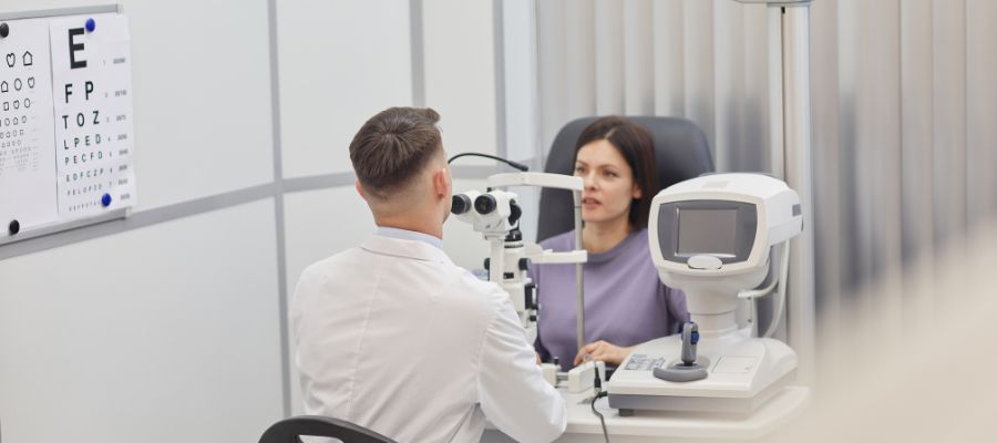
Are you in need of an advanced eye examination? Look no further than 50 Dollar Eye Guy, where Dr. Grace and Joseph Tegenkamp, along with Dr. Jeanine Spoors, provide a friendly and professional experience. With a commitment to exceptional customer service, their personalized care ensures a comfortable experience for all their patients. The doctors at 50 Dollar Eye Guy are passionate about providing high-quality care, offering Comprehensive Eye Exams and a wide selection of fashionable eyewear. Don’t wait any longer, visit their Pensacola locations today and discover the new technologies in eye examination that will enhance your vision and overall eye health.
New technologies in eye examination
In the ever-evolving field of eye care, new technologies are constantly being developed to improve the accuracy and efficiency of eye examinations. At 50 Dollar Eye Guy, we are committed to staying up-to-date with the latest advancements in order to provide the best possible care for our patients. In this article, we will explore several new technologies in eye examination that we offer at our locations in Pensacola, FL.
This image is property of lh3.googleusercontent.com.
Digital retinal imaging
Overview of digital retinal imaging
Digital retinal imaging is a cutting-edge technology that allows optometrists to capture high-resolution images of the retina, the light-sensitive tissue at the back of the eye. This non-invasive procedure provides a detailed view of the retina, including the optic nerve, blood vessels, and macula.
Advantages of digital retinal imaging
Digital retinal imaging offers numerous advantages over traditional methods of examining the retina. The high-resolution images obtained through this technology allow for early detection and monitoring of various eye diseases, such as glaucoma, macular degeneration, and diabetic retinopathy. It also provides a baseline for future comparisons, making it easier to track any changes in the retina over time.
Procedure for digital retinal imaging
During a digital retinal imaging procedure, our optometrists will use a specialized camera to capture images of the retina. The process is quick and painless, typically taking only a few minutes to complete. The images are then reviewed and analyzed by our knowledgeable optometrists.
Uses of digital retinal imaging
Digital retinal imaging has a wide range of uses in eye care. It is particularly valuable for early detection and monitoring of eye diseases, as well as for assessing the overall health of the retina. It can also be used to document any abnormalities or changes in the retina over time, assisting in the development of personalized treatment plans for patients.
Optical coherence tomography
Overview of optical coherence tomography
Optical coherence tomography (OCT) is a non-invasive imaging technique that provides detailed cross-sectional images of the retina and other structures within the eye. It uses light waves to create a 3D map of the eye, allowing optometrists to visualize the different layers of tissue.
Advantages of optical coherence tomography
OCT offers several advantages over traditional imaging techniques. It provides high-resolution images that allow for early detection and monitoring of various eye conditions, including macular degeneration, diabetic retinopathy, and glaucoma. It also enables optometrists to precisely measure the thickness of the retina, which can be useful in assessing the progression of certain eye diseases.
Procedure for optical coherence tomography
During an optical coherence tomography procedure, a specialized machine will be used to scan the eye and capture detailed images. The patient will be asked to sit still and look at a target while the scan is being performed. The process is painless and only takes a few minutes to complete.
Uses of optical coherence tomography
Optical coherence tomography is used for a variety of purposes in eye care. It is particularly valuable for diagnosing and monitoring conditions that affect the retina, such as macular degeneration and diabetic retinopathy. It can also be used to assess the integrity of the optic nerve and evaluate the success of certain eye surgeries.
Corneal topography
Overview of corneal topography
Corneal topography is a diagnostic technique that provides a detailed map of the cornea, the clear front surface of the eye. It measures the curvature of the cornea and generates a color-coded map that illustrates its shape and any irregularities.
Advantages of corneal topography
Corneal topography offers several advantages in the field of eye care. It provides valuable information for fitting contact lenses and determining the need for refractive surgeries, such as LASIK. It can also assist in the diagnosis and management of various corneal conditions, including keratoconus and astigmatism.
Procedure for corneal topography
During a corneal topography procedure, the patient’s eye will be gently touched with a special instrument that captures multiple images of the cornea. These images are then analyzed by software, which generates a detailed map of the corneal surface.
Uses of corneal topography
Corneal topography has many uses in eye care. It is essential for fitting contact lenses accurately, as it helps determine the best lens shape and size for optimal comfort and vision correction. It is also used in the pre-operative evaluation of refractive surgeries, allowing the surgeon to customize treatment plans based on the corneal curvature.
Visual field testing
Overview of visual field testing
Visual field testing is a diagnostic procedure that assesses the full extent of a person’s peripheral vision. It measures the sensitivity of the visual field and can detect any areas of decreased vision or blind spots.
Advantages of visual field testing
Visual field testing is an invaluable tool in the early detection and monitoring of various eye conditions, such as glaucoma and optic nerve damage. It helps identify any changes in the peripheral vision that may be indicative of a problem. It can also aid in the assessment of visual function, particularly in patients with neurological disorders.
Procedure for visual field testing
During a visual field testing procedure, the patient will be asked to focus on a central target while a series of lights or stimuli are presented at different locations within the visual field. The patient must indicate when they see the lights or stimuli, allowing the optometrist to map the entire visual field.
Uses of visual field testing
Visual field testing is used in the diagnosis and management of various eye conditions. It is particularly valuable for monitoring the progression of glaucoma, as well as assessing the extent of optic nerve damage. It can also be used to evaluate the impact of certain medications on the visual field and to assess visual function in patients with neurological disorders.

This image is property of s3.amazonaws.com.
Autorefractors
Overview of autorefractors
Autorefractors are automated devices used to measure a person’s refractive error, which determines their prescription for glasses or contact lenses. These devices provide quick and objective measurements of the eye’s refractive status.
Advantages of autorefractors
Autorefractors offer several advantages over traditional subjective refraction methods. They provide objective measurements, reducing the potential for errors introduced by the patient’s responses or the examiner’s interpretation. They are also faster and more efficient, allowing optometrists to perform multiple measurements in a short amount of time.
Procedure for autorefractors
During an autorefractor procedure, the patient will be asked to look into the device while it measures the focus of light reflected from the back of the eye. The measurements are then analyzed by the device, and an estimation of the patient’s refractive error is generated.
Uses of autorefractors
Autorefractors are used to determine a person’s refractive error, which is essential for prescribing the appropriate glasses or contact lenses. They are particularly useful when examining young children or individuals who have difficulty with subjective refraction techniques.
Digital refraction systems
Overview of digital refraction systems
Digital refraction systems are advanced technologies that automate the process of determining a person’s prescription for glasses or contact lenses. They offer a precise and efficient way to measure the refractive error and customize the prescription for each patient.
Advantages of digital refraction systems
Digital refraction systems provide several advantages compared to traditional manual refraction methods. They offer greater accuracy and precision, as they eliminate the potential errors introduced by manual adjustments. They also streamline the refraction process, making it quicker and more comfortable for patients.
Procedure for digital refraction systems
During a digital refraction procedure, the patient will be asked to look through a device that contains multiple lenses. The optometrist will remotely control the lenses and make precise adjustments to measure the patient’s refractive error. The patient can provide feedback on the clarity of vision, allowing the optometrist to fine-tune the prescription.
Uses of digital refraction systems
Digital refraction systems are used to determine the precise prescription for glasses or contact lenses. They are particularly valuable in cases where the refractive error is complex or when a high degree of accuracy is required. They can also be used to evaluate changes in the refractive error over time, assisting in the management of certain eye conditions.
This image is property of lh3.googleusercontent.com.
Dry eye analyzers
Overview of dry eye analyzers
Dry eye analyzers are specialized devices used to assess the quantity and quality of tears in the eyes. They provide valuable information in the diagnosis and management of dry eye syndrome, a common condition characterized by insufficient tear production or poor tear film stability.
Advantages of dry eye analyzers
Dry eye analyzers offer several advantages in the evaluation of dry eye syndrome. They provide objective measurements of tear volume, tear break-up time, and tear osmolarity, allowing optometrists to accurately diagnose and monitor the condition. They also help identify the underlying causes of dry eye and guide treatment plans.
Procedure for dry eye analyzers
During a dry eye analysis, the patient’s eyes will be examined using specialized instruments that measure tear volume, tear break-up time, and tear osmolarity. These measurements are then analyzed and used to determine the severity of dry eye and develop an appropriate treatment plan.
Uses of dry eye analyzers
Dry eye analyzers are essential tools in the diagnosis and management of dry eye syndrome. They help evaluate the effectiveness of various treatments and guide the optometrist in determining the most suitable treatment plan for each patient. They can also be used to assess the quality of tears in contact lens wearers and provide recommendations for lens selection.
Binocular indirect ophthalmoscopy
Overview of binocular indirect ophthalmoscopy
Binocular indirect ophthalmoscopy is a technique used to examine the inside of the eye, including the retina, optic nerve, and blood vessels, using a specialized handheld instrument called a binocular indirect ophthalmoscope. This procedure provides a wide-field view of the posterior segment of the eye.
Advantages of binocular indirect ophthalmoscopy
Binocular indirect ophthalmoscopy offers several advantages over other methods of examining the posterior segment of the eye. It provides a larger field of view, allowing optometrists to visualize a larger portion of the retina. It also provides a stereoscopic view, which enhances depth perception and allows for better assessment of certain eye conditions.
Procedure for binocular indirect ophthalmoscopy
During a binocular indirect ophthalmoscopy procedure, the patient will be asked to sit in a darkened room and focus on a target while the optometrist uses a headband-mounted ophthalmoscope to examine the inside of the eye. The examination is painless and generally well-tolerated by patients.
Uses of binocular indirect ophthalmoscopy
Binocular indirect ophthalmoscopy is used to assess the health of the retina, optic nerve, and blood vessels in the eye. It is particularly valuable for diagnosing and monitoring conditions such as retinal detachments, diabetic retinopathy, and macular degeneration. It can also be used to evaluate the effectiveness of certain treatments and guide surgical interventions.
This image is property of lh3.googleusercontent.com.
Slit lamp biomicroscopy
Overview of slit lamp biomicroscopy
Slit lamp biomicroscopy is a diagnostic technique that uses a high-intensity light source and a microscope to examine the structures of the eye in detail. This non-invasive procedure provides a magnified view of the anterior and posterior segments of the eye.
Advantages of slit lamp biomicroscopy
Slit lamp biomicroscopy offers several advantages in the evaluation of eye conditions. It provides a highly detailed and magnified view of the structures in the eye, allowing for precise assessment and documentation of any abnormalities. It also enables optometrists to evaluate the health of structures such as the cornea, iris, lens, and anterior chamber.
Procedure for slit lamp biomicroscopy
During a slit lamp biomicroscopy procedure, the patient will be asked to position their chin and forehead on the slit lamp instrument. The optometrist will then use a high-intensity light source and a microscope to examine the eye. The examination is painless and generally well-tolerated by patients.
Uses of slit lamp biomicroscopy
Slit lamp biomicroscopy has a wide range of uses in eye care. It is commonly used to diagnose and monitor conditions such as cataracts, corneal ulcers, and uveitis. It is also used to evaluate the fit of contact lenses and assess the success of certain eye surgeries.
Intraocular pressure measurement
Overview of intraocular pressure measurement
Intraocular pressure (IOP) refers to the fluid pressure inside the eye. Abnormalities in IOP can indicate the presence of certain eye conditions, such as glaucoma. Intraocular pressure measurement is a diagnostic procedure used to assess the pressure within the eye.
Advantages of intraocular pressure measurement
Intraocular pressure measurement is a critical component of comprehensive eye examinations, particularly in the detection and monitoring of glaucoma. Early detection of elevated IOP can help prevent vision loss and preserve the health of the optic nerve. It is also helpful in evaluating the effectiveness of glaucoma treatments and adjusting medications as needed.
Procedure for intraocular pressure measurement
There are several methods for measuring intraocular pressure, including the Goldmann applanation tonometry, air puff tonometry, and handheld tonometry devices. These methods involve gently contacting the surface of the eye or applying a gentle force to measure the resistance of the eye to indentation.
Uses of intraocular pressure measurement
Intraocular pressure measurement plays a crucial role in the diagnosis and management of glaucoma. It helps identify individuals at risk of developing the disease and allows for early intervention. It is also used to monitor the progression of glaucoma and evaluate the effectiveness of the prescribed treatments.
In conclusion, new technologies in eye examination have revolutionized the field of eye care, allowing optometrists to provide more accurate diagnoses and personalized treatment plans. At 50 Dollar Eye Guy, we are committed to incorporating these advanced technologies into our practice to ensure the best possible care for our patients. By utilizing digital retinal imaging, optical coherence tomography, corneal topography, visual field testing, autorefractors, digital refraction systems, dry eye analyzers, binocular indirect ophthalmoscopy, and intraocular pressure measurement, our experienced optometrists can detect and manage a wide range of eye conditions with precision and efficiency. If you are in the Pensacola area, we invite you to visit one of our locations and experience the benefits of these state-of-the-art technologies for yourself. Our friendly and professional team is dedicated to providing exceptional eye care and personalized service to all our patients. Make an appointment today and let us help you maintain optimal eye health and vision.
This image is property of lh3.googleusercontent.com.

